At Sandton Radiology, we provide traditional radiology services which includes plain film x-ray, ultrasound and specialised technology such as CT scans and MRI. Digital radiology is offered at Morningside with state-of-the-art DR equipment allowing for digital archiving of plain x-rays and scans. This allows for high resolution, efficient imaging limiting unnecessary radiation exposure.

We offer a full array of imaging Services, including the following:
X-Ray
X-rays are a type of radiation called electromagnetic waves. X-ray imaging creates pictures of the inside of your body. The images show the parts of your body in different shades of black and white. This is because different tissues absorb different amounts of radiation. It can help your doctor view the inside of your body without having to make an incision. This can help them diagnose, monitor, and treat many medical conditions.

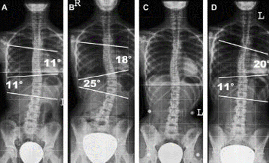
Scoliosis Studies
Scoliosis is defined as a lateral spinal curvature. Images of the entire spine is analysed for the degree of scoliosis and are measured for the purpose of correction.

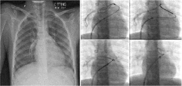
Fluoroscopy
Fluoroscopy is a low dose, real-time X-ray, used for procedures such as barium studies, angiograms, arthrograms, cystograms and interventional radiology, such as the positioning of stents and coils.
Sonar

Ultrasound imaging, also called sonography, uses sound waves to produce ultrasound images of the inside of the body. An instrument called a transducer emits high-frequency sound, inaudible to human ears, and then records the echoes as the sound waves bounce back to determine the size, shape, and consistency of soft tissues and organs. It is used to help diagnose the causes of pain, swelling and infection in the body's internal organs, joints and tendons. Doppler ultrasound is used to evaluate the arteries and veins to diagnose obstructions or blood clots also vascular malformities. Ultrasound is safe, noninvasive, and does not use ionizing radiation. Ultrasound is useful for guiding biopsy procedures.


Cross-Sectional & 3D Multi Planar Imaging
Cross Sectional Imaging is a discipline of radiology that encompasses the use of a number of advanced imaging techniques that enables imaging of the body in cross section.

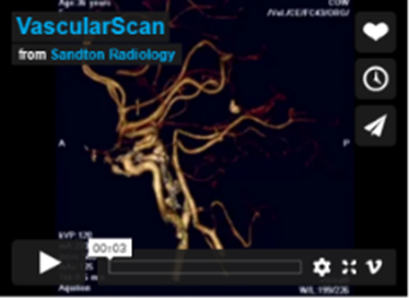
Vascular Imaging
Blood vessel imaging was previously performed with an interventional angiogram, but today CT scanning is taking angiography into a new frontier. Vessels are accurately evaluated with 3D scans performed non-invasively on an outpatient basis in just 10 minutes.

Cone Beam CT
Millions of people world wide have partial or complete edentulism. Dentures do not fully compensate patients adequately for tooth loss cosmetically or functionally. To address these issues dental implants are the accepted alternative. Dentists and oral surgeons will often initially assess their patients with x-ray. Dental CT programs offer more detailed and accurate information providing comprehensive preoperative assessment. Prior to performing this procedure, analysis of the bone of the mandible and maxilla is required in order to plan the appropriate implant procedure and limit surgical complications. The scanner allows for planning of dental implants by reconstructing the area of interest, identifying bone volume and vital nerves that need to be avoided. Excellent modality for bone detail and visualization of inner ear and paranasal sinusses. Low radiation dose especially useful in pediatrics.


MRI

In MRI the magnetic nuclei of the patient are aligned in a strong uniform magnetic field, absorb energy from tuned radiofrequency pulses, and emit radiofrequency signals as their excitation decays. This allows for multiplanar demonstration of anatomy with high resolution without the use of x-ray radiation. MRI is also used for conditions in the skull base and neck soft tissues, heart, mediastinum and aorta, abdominal and pelvic organs and for soft tissue mass assessment. Other useful applications include non-invasive vascular assessment (MR angiography), and three dimensional demonstration of structures such as the biliary tree (MRCP) and renal tract, for example. MRI is most commonly used for investigation of the brain, spine and major joints. In some cases intravenous gadolinium contrast is administered.
We have a Multi Planar cross sectional & 3D Imaging 1.5T machines at both Branches, providing no radiation and excellent soft tissue definition. We cater for claustrophobic patients and offer sedation and general anaesthesia. Please inform us if you have a pacemaker or any metallic implants.
PET/CT

This is a new advanced imaging tool that combines the modalities of CT (computed tomography) and PET (positron emission tomography) in one imaging procedure. The PET scan detects the metabolic signal of actively growing cancer cells in the body whilst the CT scan provides a detailed picture of the internal anatomy that shows the location, size and shape of cancerous growth. The results of PET/CT scans are digitally fused together providing complete information on cancer location and abnormal cellular metabolism. PET/CT combines the high sensitivity of PET with the high anatomical spatial resolution of multislice CT. This integrated approach of PET and CT technologies into a single imaging procedure permits accurate tumour detection and anatomical localisation for many cancerous growths.

